How DocLane works
From the single-slide AI inference pipeline to the multimodal integration engine.
AI inference pipeline
Whole slide image
Routine H&E slide digitized at the partner hospital.
Tiling
WSI is divided into thousands of analyzable tiles.
Foundation model
DocLane encodes morphological representations.
Feature extraction
Tile-level features aggregated to slide level.
Mutation prediction
Model scores likely genomic alterations and TMB.
Clinical decision support
Oncologist reviews AI-assisted output alongside the chart.
Each whole-slide image passes through the same six stages, from digitization to a clinician-facing recommendation — typically resolving same-day rather than in the 4–6 weeks a standard NGS turnaround requires.
Multimodal platform
Four modalities — clinical records, radiology, digital pathology and genomics — flow into a single AI engine, which outputs mutation risk, biomarker reports, therapy recommendations and resistance alerts.
The same slide your pathologist already reads
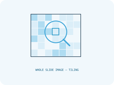
A whole-slide image (WSI) is a high-resolution digital scan of a routine glass H&E slide — the same slide already produced during standard histopathology review. No new sample collection is required.
Each WSI is divided into thousands of smaller tiles before being passed to the foundation model, since gigapixel-scale images can't be processed as a single unit by current vision architectures.
A model trained on real-world histopathology at scale
Prov-GigaPath is a self-supervised pathology foundation model, pretrained on over a billion pathology image tiles. Self-supervision means it learns general visual representations of tissue without needing millions of hand-labeled examples for every task.
DocLane fine-tunes this backbone toward Indian whole-slide image and NGS cohorts, rather than training a model from scratch — the same transfer-learning approach used in the published EGFR-prediction literature we cite below.
From pixels to a mutation probability
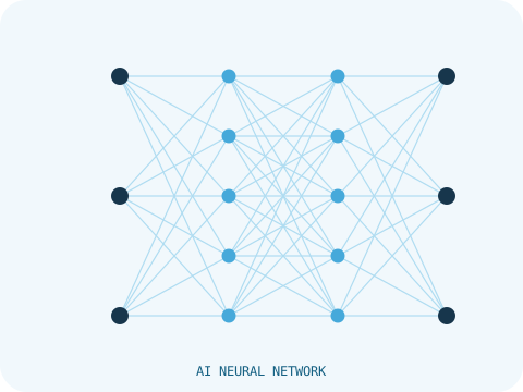
Tile-level features produced by the foundation model are aggregated into a single slide-level representation, then passed to a prediction head trained to estimate the likelihood of clinically relevant mutations and tumor mutational burden (TMB).
- Mutation risk scores for clinically actionable genes
- Tumor mutational burden (TMB) estimates
- Confidence intervals surfaced alongside every prediction
Morphology in context, not in isolation
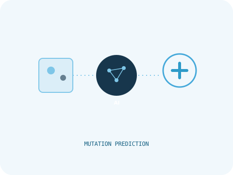
Mutation prediction from morphology alone is only one input. DocLane's multimodal layer combines it with structured clinical history, radiology and any available genomic results to produce a single longitudinal view of the patient.
The output is framed as decision support: a therapy recommendation and resistance-mechanism alert for the treating oncologist to weigh alongside their own judgment — never an autonomous prescription.